Links are not yet activated.
Is this your press release? Add a link to submitpr.org on your website and verify it — activation is instant and free.
Or upgrade with KIN for permanent activation (from $19.95).
The plate comes out of the incubator, and the clear ring looks obvious from across the bench. Then you lean in. One edge fade into a soft haze, other bulges slightly and suddenly you are no longer sure where the ring actually ends.Anyone who has worked in pharmaceutical microbiology knows this moment. It is rarely about carelessness. It is about a boundary that refuses to be simple.
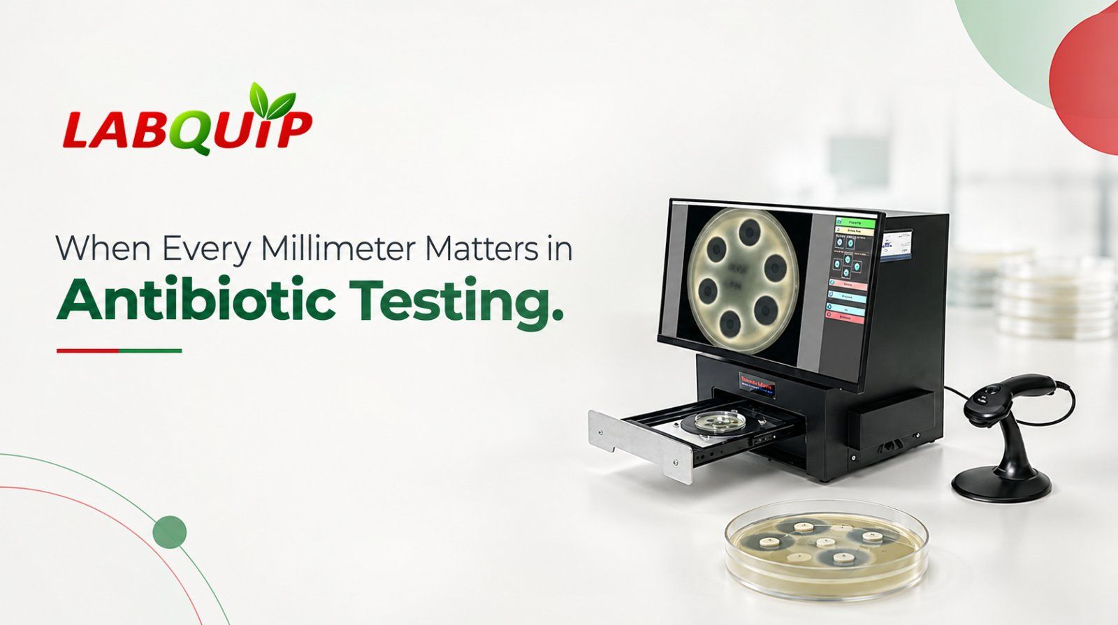
That is where an Antibiotic zone reader earns its place. By photographing the plate and measuring it with software, it gives laboratories a more structured way to handle a job that once depended on a ruler, a steady hand and trained eye. But a harder question follows: what happens when the ring itself is difficult to judge?
The honest answer depends on several things working together: the quality of the image, the nature of the ring, the software settings and the test procedure behind it. Looking at each one shows what automation can do, what it cannot promise and why it matters for antibiotic potency testing.
Challenges in Zone of Inhibition Measurement
In an agar diffusion assay, the antibiotic holds back microbial growth in the area around it. The diameter of that cleared area, called the zone of inhibition, is recorded according to the method being followed. In pharmaceutical potency testing, those diameters feed into calculations that compare the sample’s activity against a reference preparation.
Real plates are not textbook diagrams. Some rings have ragged outlines. Others fade gradually from clear to cloudy, so deciding where “inhibited” stops and “growing” begins takes careful interpretation. The organism, the agar, the incubation and the assay setup all play a part.
So, when two skilled technicians land on slightly different numbers for the same plate, neither is necessarily wrong. They are facing a genuine judgement call, and that is exactly why a consistent rule for locating the edge matters.
It also helps to separate two different problems. Trouble reading a ring is one thing. Variation created by the assay itself, such as uneven agar depth, incubation conditions or sample preparation, is another. A QC team needs to tell them apart before deciding what to fix.
A Photo Is Not a Result
Digital imaging adds a second way of looking at the plate. Instead of relying only on a ruler, the system captures the plate and presents it on screen for software-assisted measurement. Depending on the instrument, settings can be tuned to help with plates whose rings are hard to read.
But an image alone does not tell you the correct diameter. The software interprets the visible edge using its own logic, and the outcome still has to make sense for the assay in hand. Blurry rings, touching zones or unusual plate conditions deserve a second look, not automatic acceptance.
Automation can bring consistency, yet it cannot turn every ambiguous edge into a certain one. It also cannot rescue an assay that was prepared or run incorrectly. The instrument assists; the laboratory stays responsible for controlled conditions, method compliance and a proper review of the data.
This is why seeing the plate image next to the calculated diameter is so useful when choosing an Antibiotic zone reader. It lets the analyst judge at a glance whether a number looks right or needs investigating.
What a Good Antibiotic Zone Reader Should Offer
At its core, an antibiotic zone reader pairs imaging with software that measures inhibition zones and, where supported, runs the calculations for the relevant potency assay. That cuts down on repetitive measuring and manual transcription, and gives the team a clearer path for reviewing each plate.
The TRINITY V3 from Giles Scientific is built for automated zone reading and calculation in industrial antibiotic potency testing. According to the manufacturer’s documentation, it shows plate images and measured diameters on screen so users can review and confirm each size. Threshold and sensitivity settings can be adjusted to support steadier reading of difficult plates.
That matters most when a ring is not straightforward. Rather than trusting the first number the software produces, analysts can examine the image and weigh the result against the applicable method. The aim is not to remove scientific judgement but to pair it with software.
TRINITY V3 also supports potency calculations under specified pharmacopoeia methods. The manufacturer’s documentation covers USP, EP and JP, and Labquip Asia’s product page lists BP too. Included IQ, OQ and PQ documentation and support for applicable 21 CFR Part 11 electronic records and signatures are further points QA/QC teams will want to weigh. Actual compliance still depends on how each laboratory configures and uses the system.
How to Choose an Antibiotic Zone Reader
A useful instrument has to do more than display a diameter. Teams need to know how the plate image is handled, how results are shown for review, which assay calculations are covered and how records are kept for later.
Before committing, ask a few practical questions:
Does it support the assay methods we already run?
How do analysts review a tricky plate?
What documentation comes with it?
How will the equipment be qualified for its intended use?
It is equally wise to agree in advance on how unclear rings will be handled, instead of assuming every image will sort itself out.
The Takeaway
Digital imaging can make plate reading more consistent and easier to audit, but dependable results still come from three things working together: the instrument, the assay method and the people reviewing the data. The best workflow treats automation as a partner to scientific oversight, never a replacement for it.
Labquip Asia represents Giles Scientific and offers the TRINITY V3 Antibiotic Zone Reader for pharmaceutical microbiology applications. If you want to strengthen your antibiotic potency testing workflow, get in touch with Labquip Asia to discuss supported assay methods, image-review features and whether the system suits your laboratory’s needs.